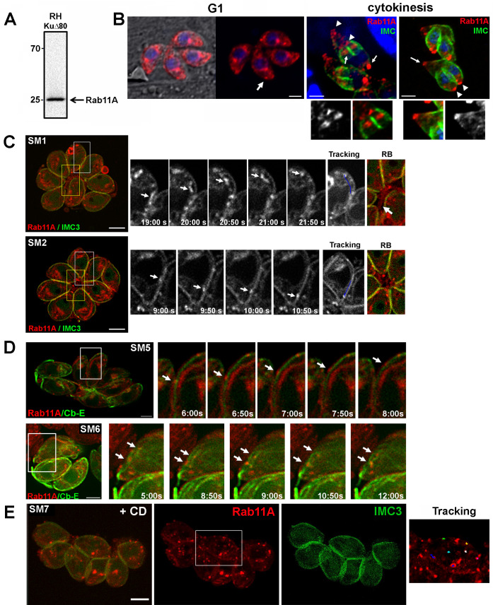
Fig 1.
A- Western blot analysis with specific anti-Rab11A antibodies detects a unique band at 25kDa in a RHΔKU80 parasite lysate. B- Analysis of Rab11A localization in fixed RHΔKU80 parasites using antibodies recognizing Rab11A, IMC3 and ROP2/3, as indicated. Bars: 1 μm. C- Sequences of images extracted from S1 Movie and S2 Movie (left images, white frames) showing the dynamic bi-directional movement of Rab11A-positive vesicles in the cytosol (upper sequence) and along the parasite cortex (lower sequence) of mcherryRab11A-WT and IMC3-YFP expressing parasites. Tracking of vesicle trajectory is also shown. Images on the right show a zoom of the residual body (RB) region indicated by a yellow frame in the corresponding vacuoles. The arrow indicates Rab11A-positive tubular-like structures connecting the parasites Bars: 2 μm. D- Sequences of images extracted from S5 Movie and S6 Movie (left images, white frames) showing the dynamic movement of Rab11A-positive vesicles along the parasite cortex (upper sequence) and in the cytosol (lower sequence) of mcherryRab11A-WT and Cb-Emerald GFP (Cb-E) expressing parasites. Bars: 2 μm. E- Images extracted from movie S7 Movie. mcherryRab11A-WT and IMC3-YFP expressing parasites were treated with cytochalasin D (CD) for 30 min before being recorded. Rab11A-positive vesicles are detected in clusters displaying confined trajectories (right image: “tracking”). Bar: 2 μm.

