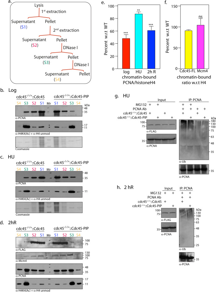
Fig 6. Cdc45 helps recruit/stabilize PCNA-polymerase complexes on chromatin during active DNA replication.
a-e. Analysis of soluble and chromatin-bound protein fractions isolated from cdc45-/-/+::Cdc45 and cdc45-/-/+::Cdc45-PIP cells. Each experiment was carried out thrice. One data set for each is presented here. Reactions resolved on 12% SDS-PAGE were probed with various antibodies as indicated. S1, S2 –soluble fractions; S3, S4 –DNA-associated fractions. Mr- molecular weight marker a. Outline of fractionation scheme b. Isolation from 5 x10⁷ logarithmically growing cells. c. Isolation from 5 x10⁷ HU treated cells d. Isolation from 1–2 x10⁹ synchronized cells 2 hours after release from block. e. ratio of chromatin-bound PCNA (S3+S4): histone H4 (S3+S4) in cdc45-/-/+::Cdc45-PIP cells, was plotted as a percentage of ratio of chromatin-bound PCNA (S3+S4): histone H4 (S3+S4) in cdc45-/-/+::Cdc45 cells. Quantification was carried out using ImageJ software. Bars represent average of three experiments, with error bars indicating standard deviation. Student's t-test was applied for statistical significance. p value <0.05*, <0.005**, <0.0005*** f. ratio of chromatin-bound Mcm4 (S3+S4): histone H4 (S3+S4) in cdc45-/-/+::Cdc45-PIP cells, was plotted as a percentage of ratio of chromatin-bound Mcm4 (S3+S4): histone H4 (S3+S4) in cdc45-/-/+::Cdc45 cells. Similar analysis was also done for chromatin-bound Cdc45-FLAG. Quantification was carried out using ImageJ software. Bars represent average of three experiments, with error bars indicating standard error of mean. Student's t-test was applied for statistical significance. ns: not significant. g-h. Analysis of PCNA immunoprecipitates from whole cell extracts of cdc45-/-/+::Cdc45 and cdc45-/-/+::Cdc45-PIP cells. g. Cells were treated with 5 mM HU for 8 hours, with 20 μM MG132 being added 5 hours into the treatment. h. Cells were harvested 2 hours after release from HU-induced block. 20 μM MG132 was added 5 hours into the HU treatment and also added to the cells at the time of release from HU. The immunoprecipitates were resolved on SDS-PAGE and analysed for ubiquitinated PCNA using anti-Ub antibodies (Santa Cruz, 1:1000 dil). The blot was subsequently probed with anti-PCNA antibody (1:2500 dil).

