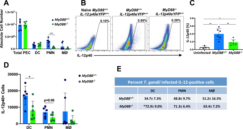
Fig 2. Multiple cell types produce MyD88-independent IL-12 during in vivo infection.
IL-12 reporter mice on a wild type (MyD88+/+IL-12p40eYFP/eYFP) and MyD88 knockout (MyD88-/-IL-12p40eYFP/eYFP) background were infected by intraperitoneal injection with 103 RH strain tachyzoites. On day 5 post-inoculation, peritoneal exudate cells were collected for ex vivo analysis. (A) Number of PEC, dendritic cells (DC), neutrophils (PMN) and macrophages (MØ) present in the peritoneal cavity. (B) Percentage of IL-12eYFP positive cells in the peritoneal cavity of one representative infected MyD88+/+IL-12p40eYFP/eYFP, one representative infected MyD88-/-IL-12p40eYFP/eYFP, and one representative noninfected MyD88+/+IL-12p40eYFP/eYFP mouse. (C) Percent IL-12 positive cells in the peritoneal cavities of WT and MyD88 KO IL-12 reporter mice. (D) Number of IL-12 positive cells amongst DC, PMN and MØ isolated from MyD88+/+ and MyD88-/- reporter mice. (E) Percent of infected cells that are positive for IL-12. In these experiments, DC were defined as MHCII+, CD11c+, F4/80- cells; PMN were defined as Ly6G+ cells; MØ were defined as F4/80+ cells. Values are the means ± SEM of 2 pooled independent experiments, n = 6 per group. Each symbol represents an individual mouse. Statistical significance was assessed using Student's t test (* p < 0.05, ** p < 0.01, *** p < 0.001) (A, C and D). Mann-Whitney test was used to determine statistical significance of IL-12-positive MyD88 WT vs. KO DC in E. These experiments were performed three times with similar results.

