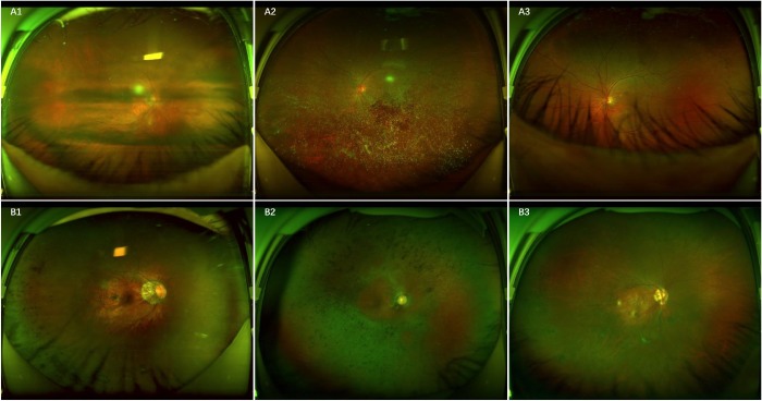
Figure 6.
Ultra-widefield fundus images showing typical false-negative and false-positive cases in RH detection. A, False-negative images: A1, scattered RH under the obscured optical media; A2, RH in the center, partly covered by the opaque vitreous body; A3, RH at the bottom, partly covered by eyelashes. B, False-positive images: B1, retinal pigmentation on the left side; B2, retinal pigmentosa; B3, round retinal holes on the left side.

