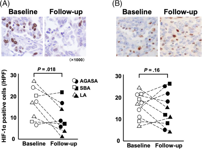
FIGURE 5.

HIF‐1α+ cells were assessed by immunohistochemistry in the tumor specimens collected at first and second excision (Figure 4 showed small bowel adenocarcinoma). In dogs receiving surgery and toceranib phosphate, the median number of HIF‐1α+ tumor cells at baseline and follow‐up were 17.8 and 8.5 cells per high power field (HPF), respectively; there was a significant decrease at follow‐up when compared to baseline (P = .02; A). In dogs receiving surgery alone, the median of baseline and follow‐up were 17.1 and 14.4 cells per HPF, respectively; there was no significant difference between baseline and follow‐up (P = .16; B)
