
FIGURE 1.
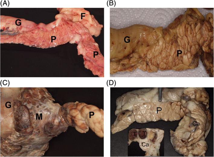
Macroscopic appearance of the pancreas in the different groups: A, normal, smooth, light red pancreas (P), gut (G), and smooth white fat tissue (F) (9‐year‐old Labrador, group 1; unfixed tissue), B, pancreatic tissue (P) with prominent lobular structure and histologically mild acute interlobular inflammation and part of the gut (G) (14‐year‐old West Highland White Terrier, group 2; formalin‐fixed tissue), C, focal severe acute purulent‐necrotising inflammation of the pancreas (P) appears grossly as a firm dark‐brown mass (M) close to the gut (G) (3‐year‐old Labrador, group 3; formalin‐fixed tissue), D, the acinar pancreatic carcinoma (Ca) is a firm mass within the pancreas (P). Inset: cut surface with homogenous white firm carcinoma mass (Ca) and thrombosed vessels (T) (5‐year‐old Flat‐Coated Retriever, group 4; formalin‐fixed tissue)