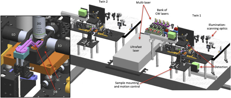
FIG. 2.
3D opto-mechanical model of the twin-microscope system mounted on a 5 × 10 ft2, anti-vibration optical table. Model shows the multi-laser subsystem shared between microscope-twin-1 (right) and microscope-twin-2 (left). Twin-1 has the four functional subsystems labeled and features an implementation of both upright and inverted detection. Brief descriptions of each functional subsystem are provided in Table I. The inset shows a detailed view of the sample chamber (SC), the dive bar (DB) used to hold the sample, three excitation objectives (EO) to deliver excitation light-sheets to the sample, and the detection objective (DO) to collect emitted fluorescence from the sample.

