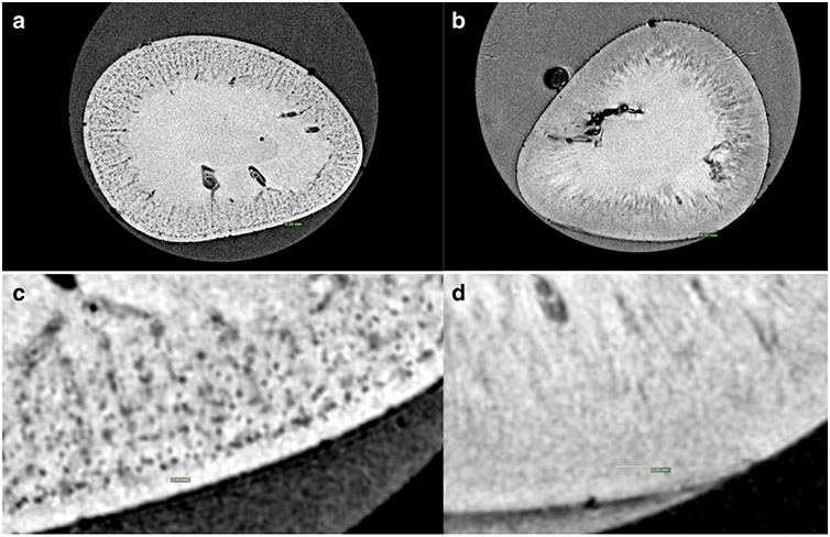
Figure 2. Representative MR image of cationic ferritin enhanced MRI (A) and control without cationic ferritin (B).
The systemically administered cationic ferritin transiently binds to the glomerular basement membrane and allows each glomerulus to be MRI detectable (a). However, even at high field strength there is not enough contrast between the glomeruli and surrounding vasculature and tubules to detect the glomeruli without an exogenous contrast agent (b). Distinct black dots can be seen in greater detail at high magnification (c) but no glomeruli are visible without the contrast (d). Green scale bar represents 0.5 mm.

