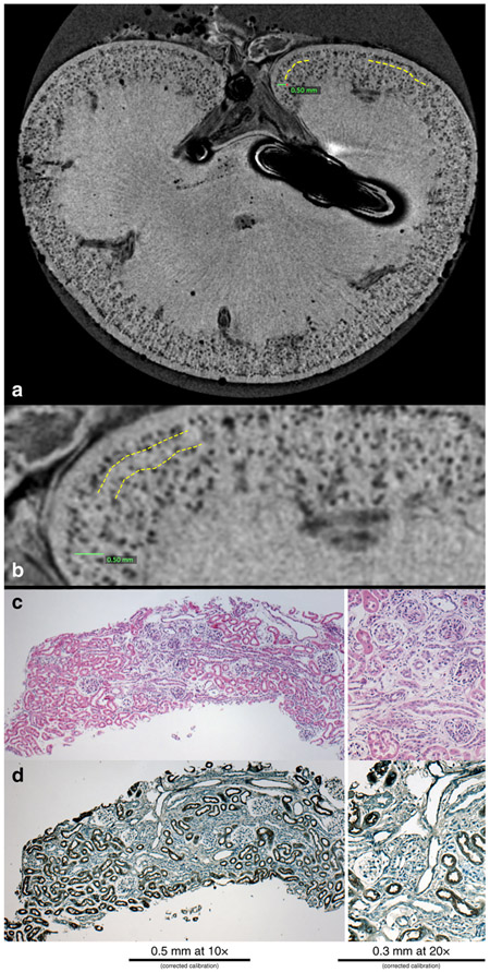
Figure 3. Nephrotoxin-induced AKI during nephrogenesis results in a circumferential layer of glomeruli where cationic ferritin is heterogeneously detected.
A representative MR image from a kidney from the AKI group where glomerular drop out is outlined in the yellow dotted lines (a). This area is magnified in (b) where the yellow dotted lines outline the boundaries of the area with glomerular drop out and there are glomeruli just under the capsule that are normal appearing and label with CF. Histologically this area has shrunken, immature glomeruli by PAS (c), with a lack of surrounding proximal tubules by lotus (d).

