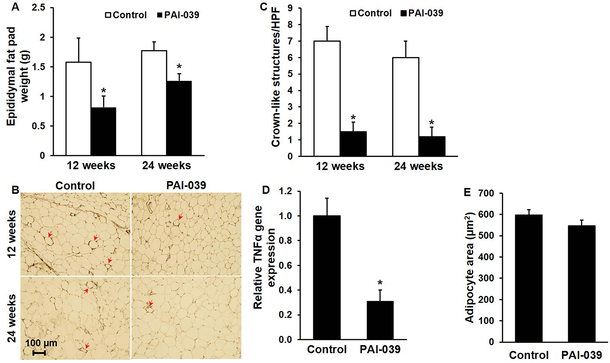
Figure 4. PAI-039 decreases visceral white adipose tissue mass and inflammation.

Epididymal white adipose tissue of ldlr−/− mice was harvested and analyzed after 12 and 24 weeks of WD with or without PAI-039. (A) Mean combined weights of left and right epididymal fat pads; n=5–7 mice/group; *P<0.05 vs. control. (B) Macrophage (Mac-3) immunostaining, demonstrating peri-adipocyte crown-like structures (arrows). (C) Quantification of crown-like structure formation; n=4–5/group, *P<0.01. HPF, high-power field. (D) TNF-α gene expression, assessed by real-time RT-PCR analysis after 24 weeks of WD; n=3–5 mice/group; *P<0.01 vs. control. (E) Mean adipocyte size (cross-sectional area), assessed after 12 weeks of WD; n=4–5/group; P>0.3.
