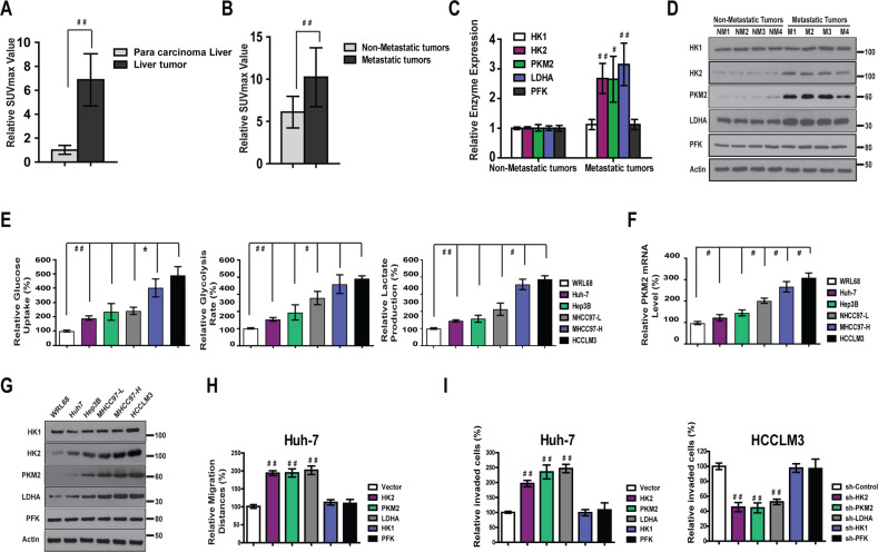
Fig. 1. The level of metabolism is positively correlate with metastatic potential of HCC.
a The levels of Suvmax (n = 121) value in were significantly higher in liver tumor than in para carcinoma tissue. b Suvmax levels were significantly higher in metastatic liver tissue (n = 32) than in non-metastatic HCC tissue (n = 84). c, d The mRNA and protein expressions of HK2, PKM2, and LDHA were much higher in HCC tumors that had more metastatic potential. e Relative glucose uptake, the rate of glycolysis and lactate production were higher in five HCC cell lines compared with normal liver cell line WRL68. f Relative PKM2 mRNA level was higher in five HCC cell lines compared with normal liver cell line WRL68. g Protein levels of HK2, PKM2, and LDHA were much higher in five HCC cell lines compared with normal liver cell line WRL68. h Overexpression of HK2, PKM2, and LDHA markedly increased wound healing capacity in Huh-7 cells. i Overexpression of HK2, PKM2, and LDHA markedly increased Matrigel invasion in Huh-7 cells. On the contrary, knockdown of HK2, PKM2, and LDHA block Matrigel invasion in HCCLM3 cells.

