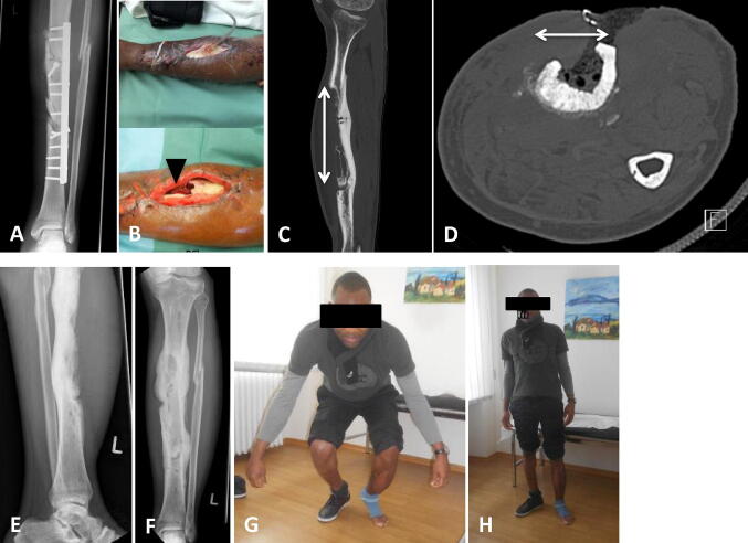
Fig. 3.
FRI after a heavy vehicle accident with a II° open tibial shaft fracture and initial fixation in Africa (a). Admission from foreign clinic after several debridements and NPWT treatments. A soft tissue (arrow mark in b) and bone (arrows in c, d) defect of around 18 cm was seen anterior to the tibial shaft. The tissue samples were positive for infection with Staphylococcus epidermidis, Pseudomonas and Escheria coli. After infection and dead space management (with Cerament™ G), the reconstruction management was completed with a local free flap (M. latissimus dorsi). Presentation 12 months after surgery with good bone consolidation and full weight-bearing (e–h)

