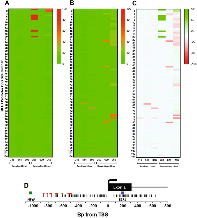
Fig. 5.
Effect of DAC treatment on MLH1 promoter methylation in three sensitized and three desensitized GBM cell lines. a Long-read SMRT-seq of bisulfite-converted amplicons of a 2.5 kb segment of the MLH1 promoter was used to quantify the percentage of methylated reads at 104 consecutive CpG sites. Compared to TMZ sensitized lines, desensitized lines showed higher levels of baseline methylation in the region containing CpGs 1–20. b Percentage of methylated reads after treatment with DAC 100 nM/days × 7 days. c Absolute change in percentage of methylated reads after DAC treatment, with demethylation depicted in green and hypermethylation depicted in red. d Schematic of the genomic region containing the CpG island at the MLH1 promoter, showing the locations of CpG sites (vertical lines), hypermethylated cytosines in desensitized lines (red), and predicted nearby transcription factor binding sites (squares) in relation to the MLH1 transcription start site (TSS). Genomic coordinate of the TSS is chr3:36,993,350 (hg38)

