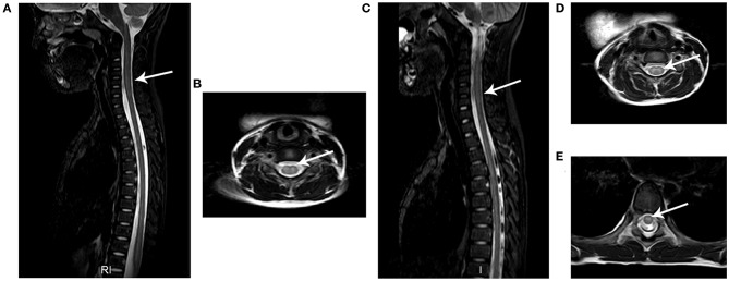
Figure 3.
Typical MRI results of AFM patients. The images from two AFM patients (case a: A,B; case b: C,E) after onset of neurological symptoms. (A,C) Sagittal T2-weighted images showing longitudinal hyperintensity (arrow) in central gray matter. (B,D,E) Axial T2-weighted sequences showing hyperintensity in spinal cord gray matter.

