Figure 2.
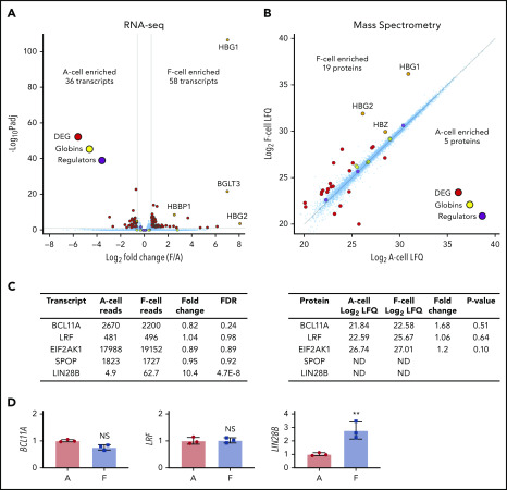
Analysis of HUDEP2-derived F and A erythroblasts. (A) RNA-seq analysis of day 7 differentiated HUDEP2 F and A cells from 3 independent subclones. False discovery rate (FDR) <0.05; fold change >1.5. (B) MS analysis of day 6 differentiated HUDEP2 F and A cells. Approximately 6000 proteins were detected. P < .05; fold change >2. Globin transcripts and known HbF regulators (BCL11A, LRF, EIF2AK1, and SPOP) are highlighted. (C) Transcript (left) and protein (right) levels of selected HbF regulators. Transcript data are shown as normalized reads. (D) RT-PCR quantification of selected HbF regulators from sorted HUDEP2 day 7 F and A cells. Data are shown as mean ± SD. **P < .01 vs A cells. DEG, differentially expressed genes; LFQ, label-free quantification intensity; Padj, adjusted P value.

