
Figure 1.
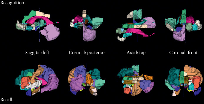
MRI regions selected for prediction of executive functions decline. For the combination with EEG during recognition and neuropsychology, volumetry from the following regions was selected as features: amygdala right, caudate nucleus right, medial, lateral, and posterior orbital gyrus left, superior temporal gyrus middle part right, bilateral lateral ventricle excluding temporal horn, subcallosal area left, bilateral pre-subgenual frontal cortex left, cerebellum right, superior temporal gyrus anterior part right, and anterior and posterior cingulate gyrus right. For the combination with EEG during recall without neuropsychology, volumetry from the following regions was selected as features: bilateral amygdala, anterior temporal lobe lateral part left, parahippocampal and ambient gyrus right, superior temporal gyrus middle part right, fusiform gyrus right, bilateral insula, lateral remainder occipital lobe left, anterior cingulate gyrus right, bilateral posterior cingulate gyrus, middle frontal gyrus right, bilateral caudate nucleus, bilateral nucleus accumbens, thalamus right, corpus callosum, lateral ventricle excluding temporal horn left, inferior frontal gyrus left, postcentral gyrus left, superior parietal gyrus left, cuneus right, posterior orbital gyrus left, substantia nigra left, subgenual frontal cortex left, subcallosal area right, and bilateral pre-subgenual frontal cortex L.