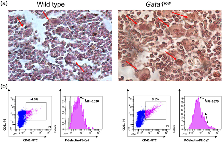
FIGURE 3.
Gata1low megakaryocytes express levels of TGF-β and of P-selectin greater than normal. (a) TGF-β-specific immunohistochemistry of bone marrow sections from representative wild-type and Gata1low littermates, as indicated. To be noted the intense immunostaining of the Gata1low megakaryocytes. Magnification ×40. Megakaryocytes are indicated by arrows. Similar results were published in Reference 44. (b) Flow cytometry analyses for the expression of P-selectin by megakaryocytes from wild-type and Gata1low littermates. Megakaryocytes were identified on the basis of CD41/CD61 markers, as indicated. Expression of high levels of P-selectin in Gata1low megakaryocytes was also detected by electron microscopy31

