Figure 3.
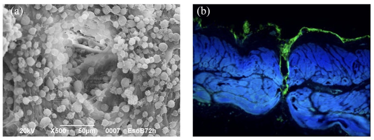
SEM image of a decellularized scaffold seeded with MSCs and cell culture outside and inside channels.
(a) SEM image of a decellularized scaffold seeded with MSCs: magnification 500×, scale bar 50 μm; (b) cell culture (actina marchers) outside and inside channels.
MSC, mesenchymal stromal cell; SEM, scanning electron microscopy.

