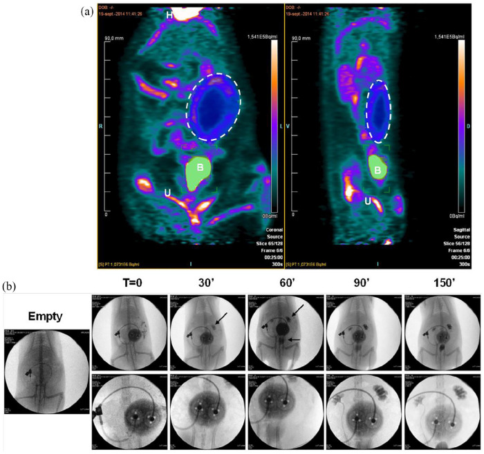
Figure 4.
Pre-vascularization of the device allows efficient exchanges with blood circulation: (a) PET-scan performed 2 days after injection of 4000 rat islets in the MailPan® after 3 months post-implantation in the peritoneal cavity of non-diabetic rats. Picture shows signal accumulation in the area of MailPan® device indicating the entry of glucose. Representative pictures of n = 3 experiments, dotted circle highlight MailPan®’s position. H: heart, B: bladder, U: urinary tract and (b) injection of angiography contrast product in MailPan® in the peritoneal cavity of rats with a 150 min follow-up. Arrows highlight the accumulation of contrast product in kidney starting 30 min and in kidney and bladder starting 60 min. Upper panel show view of the whole abdomen and lower panel a close-up of MailPan® device. Representative pictures of n = 3 experiments.

