Figure.

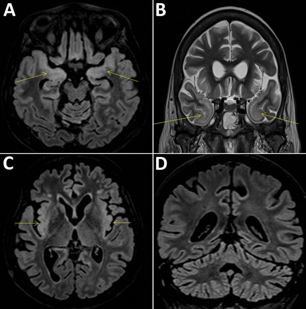
Cerebral magnetic resonance imaging scans compatible with the diagnosis of encephalitis in a 58-year-old woman, France. Fluid-attenuated inversion recovery (FLAIR) and T2 hypersignals in limbic system structures, including both amygdalae (A, arrows), temporal poles (B, arrows), and insular cortex (C), associated with FLAIR hyperintensities of the cerebellar cortex (D).
