Figure 2.
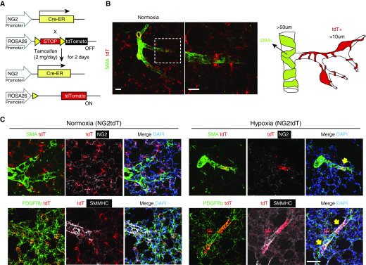
Muscularization of distal arterioles with hypoxia in NG2tdT murine line. (A) Diagram showing the strategy for generation of NG2tdT mice. (B) The landscape of the NG2tdT lung with diagram. The dashed box represents the higher magnification of the area. Scale bars: 50 μm. (C) Left lung of normoxia and hypoxia NG2tdT was stained for SMA (SMC marker, green), NG2 (mural cell marker, white), PDGFRb (pericyte and SMC marker, green), SMMHC (SMC marker, white), and nuclei (DAPI, blue). Note that tdTomato (red) is a reporter color and marked with the lineage tag without antibody labeling. The yellow arrows indicate tdT+ cells cover areas of a small arteriole and are PDGFRb+SMMHC+. Scale bar: 50 μm. ER = estrogen receptor; tdT = tdTomato.

