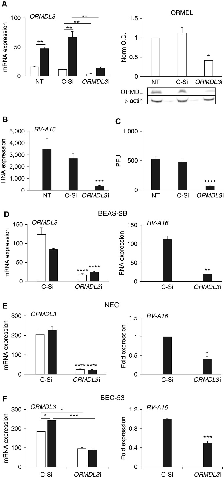
Figure 2.
ORMDL3 supports RV-A16 replication in human epithelial cells. Open bars: uninfected. Solid bars: RV-A16 infected. (A) ORMDL3 expression and protein: HeLa cells were transfected with control (C-Si) or ORMDL3 siRNA (ORMDL3i) 24 hours before infection for 48 hours. Cells were processed for qPCR and data normalized to 18S rRNA (left; N = 5). Right: cell lysates from siRNA-transfected cells were analyzed for ORMDL protein by Western blot. ORMDL optical density (O.D.) was normalized to β-actin O.D. (right; N = 3; P < 0.05 versus untreated [NT] and control siRNA). (B) Samples from A were analyzed by qPCR for RV-A16 RNA with normalization to 18S rRNA. Results are from five independent experiments. (C) A plaque-forming unit (PFU) assay from siRNA-transfected cells after 48 hours of infection. N = 3 independent experiments. P < 0.001 compared with NT or C-Si cells. (D) BEAS-2B cells were knocked down for ORMDL3, infected with RV-A16 for 48 hours, and processed as above. qPCR results were normalized to 18S rRNA. N = 4 independent experiments. (E) Primary nasal epithelial cells (NECs) were knocked down for ORMDL3 and then infected with RV-A16 for 48 hours. RNA was processed for qPCR. RV-A16 RNA was normalized to C-Si samples (set = 1). The experiment was repeated with four individual donors. (F) Primary bronchial epithelial cells from one donor (BEC-53) were knocked down for ORMDL3, infected with RV-A16 for 48 hours, and processed and normalized as in E. N = 3 independent experiments using cells from this donor. *P ≤ 0.05, **P ≤ 0.01, ***P ≤ 0.005, and ****P ≤ 0.001.

