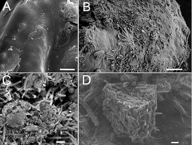
Figure 5. SEM images of cell cultures of primmorphs.
(A) The epithelial surface of healthy cultures was clean, flat and smooth. (B) The surface of the primmorphs infected with the cellular suspension from the diseased sponge. The melting of the sponge epithelial cells and increase in different bacteria on day 7 were observed. (C) The primmorphs infected with cellular suspension from the diseased sponge, the death of green algae symbionts, sponge cells and massive growth of different bacteria for 21 day. (D) Bio-cake formed in infected cultures of primmorphs from diseased sponge on day 30. Scale bars are 1 µm.

