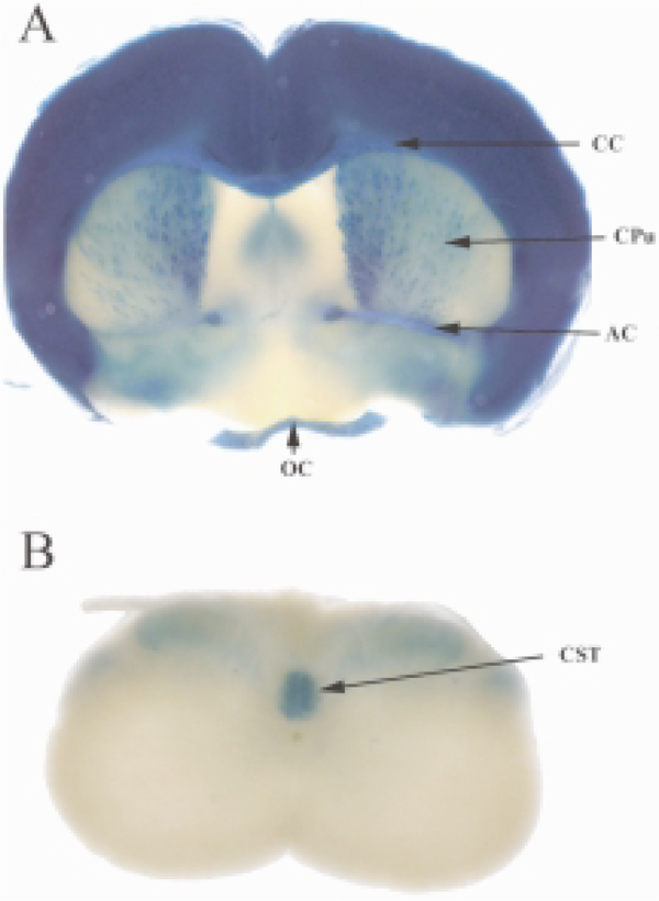
Fig. 3.
NgR1 expression in brain and spinal cord. β-galactosidase reporter gene expression analysis in NgR1tauLacZ mice (Zheng et al., 2005), as assessed by X-gal histochemistry. A. In brain tissue sections of one-month-old mice, strongest β-galactosidase activity is observed in the cerebral cortex, the corpus callosum (CC), anterior commissure (AC), and optic chiasm (OC). Weaker β-galactosidase activity is found in the medial septum and the striatum/caudate putamen (CPu). B. In the spinal cord of one-month-old mice, β-galactosidase activity is most robust in the corticospinal tract (CST). Weaker labeling is detected in the dorso-lateral spinal while matter including presumptive fiber tracts such as the rubrospinal or raphespinal tract. Also weak labeling is detected in the dorsal gray matter of the spinal cord, and may arise from DRG neuron afferent projections. No reporter gene expression was detected in motoneuron pools or the ventral gray and white matter of the spinal cord. This suggests that robust NgR1 expression in the spinal cord is restricted to a small number of fiber tracts.

