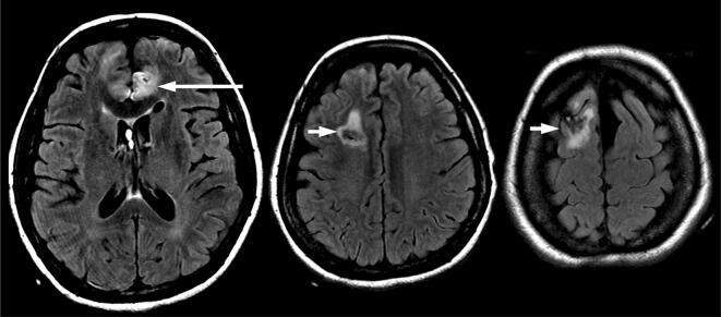
Fig. 4.
MRI brain 3 weeks after presentation. Axial T2-weighted FLAIR images show the small infarct in the left anterior cerebral artery territory (long arrow) which was already present 8 days after presentation, and edema around the right-sided ventricular drainage catheter tract (short arrows), but no subsequent infarction in the anterior cerebral artery territories

