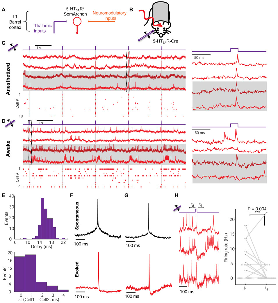
Figure 2. Sensory-evoked responses in L1 neurons in vivo.
(A) 5-HT3AR-positive interneurons in barrel cortex L1 receive thalamocortical (sensory) and neuromodulatory inputs. (B) Simultaneous sensory stimulation and voltage imaging in L1. (C) Fluorescence transients in single L1 interneurons evoked by whisker stimuli (20 ms deflections at 0.5 Hz) in isoflurane anesthetized mice. Left, top: example fluorescence traces. Horizontal stripes indicate simultaneously recorded cells. Bottom: spike raster (n = 18 neurons, 3 mice). Right: Fluorescence waveforms from the boxed region at left. (D) Same as C but in awake mice. (E) Top: Distribution of delays between stimulus onset and peak of evoked spike (n = 24 cells, 135 trials). Bottom: Distribution of relative delays in sensory-evoked action potential peaks between simultaneously recorded pairs of cells (n = 14 pairs, 66 trials). (F) Spike-triggered average waveform of spontaneous (top, n = 17 neurons) and whisker stimulus-evoked (bottom, n = 24 neurons) action potentials. (G) Same as F but in an awake mouse. Spontaneous: n = 22 neurons. Evoked: n = 21 neurons, 3 mice. Stimulus-evoked spikes had smaller after-spike hyperpolarization under anesthesia than under wakefulness (anesthetized: 11 ± 2% spike height, n = 24 neurons, 3 mice vs. awake: 17 ± 1% of spike height, n = 21 neurons, 3 mice, p = 0.02, two-tailed t-test), consistent with a more depolarized resting potential under wakefulness (Constantinople and Bruno, 2011).(H) Sensory stimulation induced a period of reduced spontaneous activity in awake mice.

