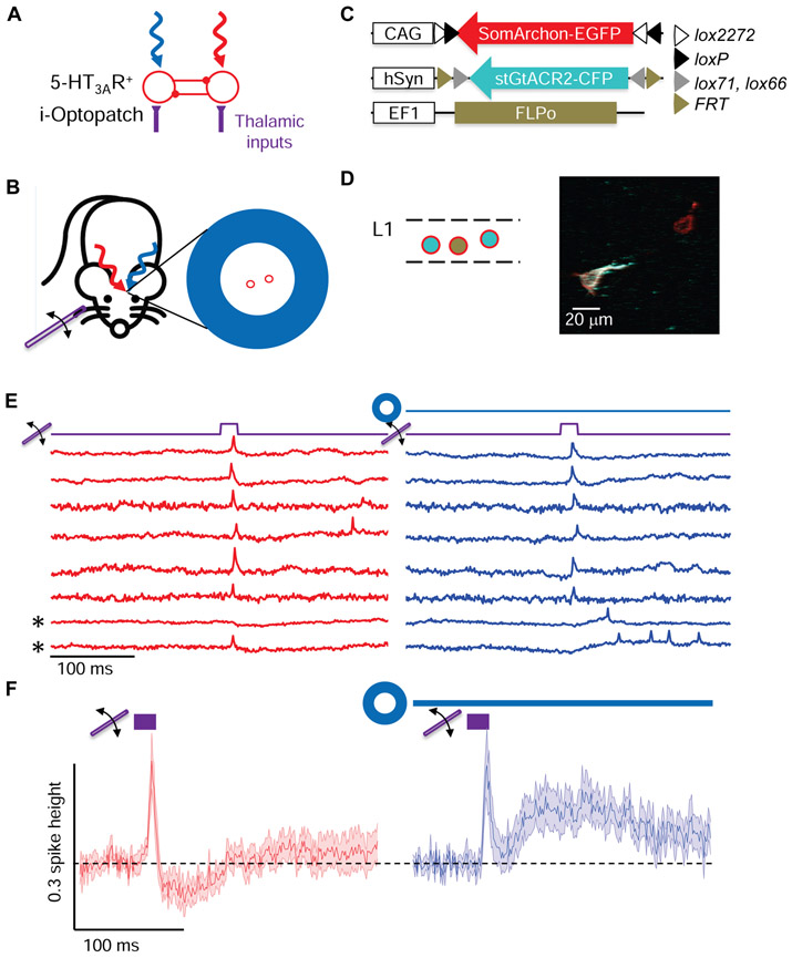
Figure 5. Optogenetic silencing reveals that L1 network activity is necessary for sensory-evoked lateral inhibition.
(A,B) Sensory-evoked responses in centrally located neurons were recorded (red) either without or with optogenetic silencing of surrounding L1 neurons (blue). (C, D) Targeted gene expression. Cre-on SomArchon-EGFP expressed in all 5-HT3AR-Cre+ neurons. Cre-on-flp-off stGtACR2-CFP and low-titer Flpo virus were combined, so stGtACR2-CFP expressed in Cre+/Flp− neurons. (D) Left: schematic of gene expression patterns. Colors indicate constructs in (C). Right: Composite two-photon fluorescence image of GFP fluorescence from SomArchon-EGFP and CFP fluorescence from stGtACR2-CFP. Voltage imaging was performed only in neurons that did not express stGtACR2. (E) Paired recordings in awake mice of sensory-evoked responses from individual centrally located neurons, either without (red) or with (blue) inhibition of surrounding neurons. Asterisks indicate recordings where sensory stimuli evoked delayed spiking when lateral inhibition was suppressed. (F) Whisker stimulus-triggered average membrane potential without (red) or with (blue) surround inhibition (n = 13 neurons, 2 mice, shading represents s.e.m.).

