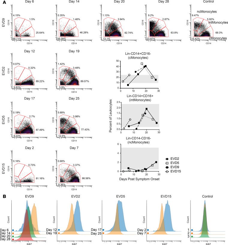
Figure 3. Monocyte populations are lost and then recover during EVD.
(A) Loss of intermediate and nonclassical monocytes during the acute viremic phase followed by recovery, of first intermediate and then nonclassical monocytes, in convalescence. Quantitation of these cell populations as a percentage of CD45+CD66– leukocytes is shown in graphs (range for all controls in gray). (B) CD14+ monocytes are actively proliferating (Ki-67+) during the acute phase at time points in which viral loads are declining. Samples from a control obtained on 3 separate dates are shown for comparison.

