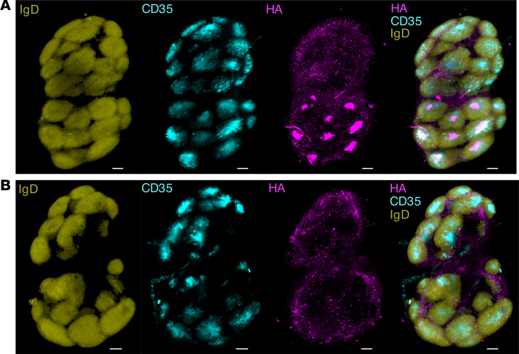
Figure 4. Increased deposition of HA-ferritin in GCs.
C57BL/6 mice (n = 2 mice per group) were immunized with Alexa Fluor 647–labeled HA-ferritin (5 μg) (A) or a molar equivalent of Alexa Fluor 647–labeled soluble HA (3.8 μg) (B), adjuvanted with AddaVax. After 14 days, draining inguinal LNs were stained (IgD, to identify B cells, yellow; CD35, a marker of FDCs, blue), cleared and imaged by lightsheet microscopy. Images are maximum intensity projections of Z-stacks and are representative of each treatment group. Scale bar: 200 μm.

