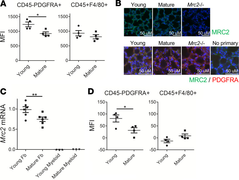
Figure 2. Fibroblasts exhibit decreased Mrc2 expression during maturation.
(A) Fluorescent collagen uptake assay in lung cells after gating for markers as indicated; n = 4 female mice in each group. (B) Representative immunofluorescence staining of tissue sections from WT mouse lungs with sections from Mrc2–/– mice serving as control; top: original magnification, ×400, with MRC2 in green; bottom: original magnification, 630×, with MRC2 in green, costained with PDGFRA in red; no primary, no PDGFRA antibody; DAPI is a counterstain. (C) Q-RT-PCR in sorted fibroblasts or myeloid cells for Mrc2; n = 3–6;a mix of male and female mice were used. (D) Flow cytometric staining for MRC2 after gating for markers as indicated; n = 4; a mix of male and female mice were used. Statistics: (A and D) Student’s t test, (C) ANOVA. *P < 0.05, **P < 0.01.

