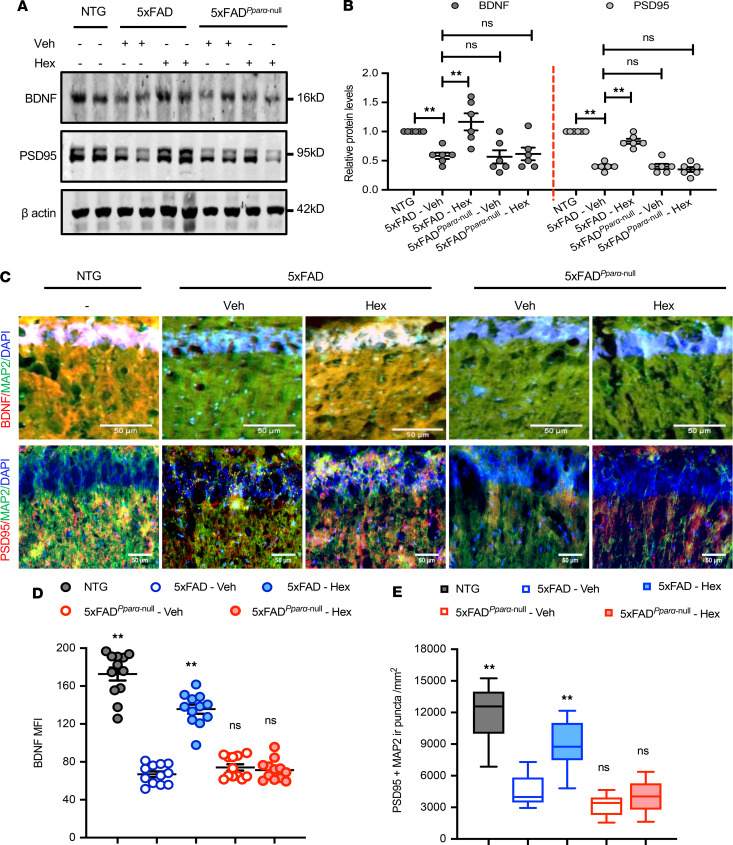
Figure 5. Hex upregulates BDNF and PSD95 in the hippocampi of 5XFAD mice via PPARα.
(A) 5XFAD and 5XFADPparα-null mice (n = 6/group; 6–7 months old) were treated orally with Hex (5 mg/kg/d) or vehicle (0.1% methyl cellulose) for 1 month followed by analysis of BDNF and PSD95 proteins by immunoblot. Uncropped Western blot images are shown in Supplemental Figure 6. (B) Relative density of BDNF and PSD95 protein expressions compared with β-actin was calculated by ImageJ. Results are shown as mean ± SEM. One-way ANOVA followed by Bonferroni’s multiple comparisons test was used to assess the significance of the mean; **P < 0.01 vs. vehicle-fed 5XFAD, nsP > 0.05 vs. vehicle-fed 5XFAD. (C) Hippocampal sections were double labeled with either BDNF (red) plus MAP2 (green) or PSD95 (red) plus MAP2 (green). Scale bar: 50 μm. Raw images are shown in Supplemental Figure 6. (D) MFI of hippocampal BDNF was quantified in 2 sections of each of 6 mice per group. One-way ANOVA [BDNF MFI: F(4,55) = 114.28, P < 0.001], followed by Bonferroni’s multiple comparisons test was used; **P < 0.01 vs. vehicle-fed 5XFAD, nsP > 0.05 vs. vehicle-fed 5XFAD. (E) Colocalization of PSD95 with MAP2 was analyzed by quantifying PSD95 and MAP2 immunoreactive puncta using ImageJ. For quantification, 2 sections per mice (n = 6 per group) was used. Results are shown as mean ± SEM. One-way ANOVA [PSD95 puncta: F(4,55) = 52.569, P < 0.001], followed by Bonferroni’s multiple comparisons test was used to assess the significance of the mean, **P < 0.01 vs. vehicle-fed 5XFAD, nsP > 0.05 vs. vehicle-fed 5XFAD.

