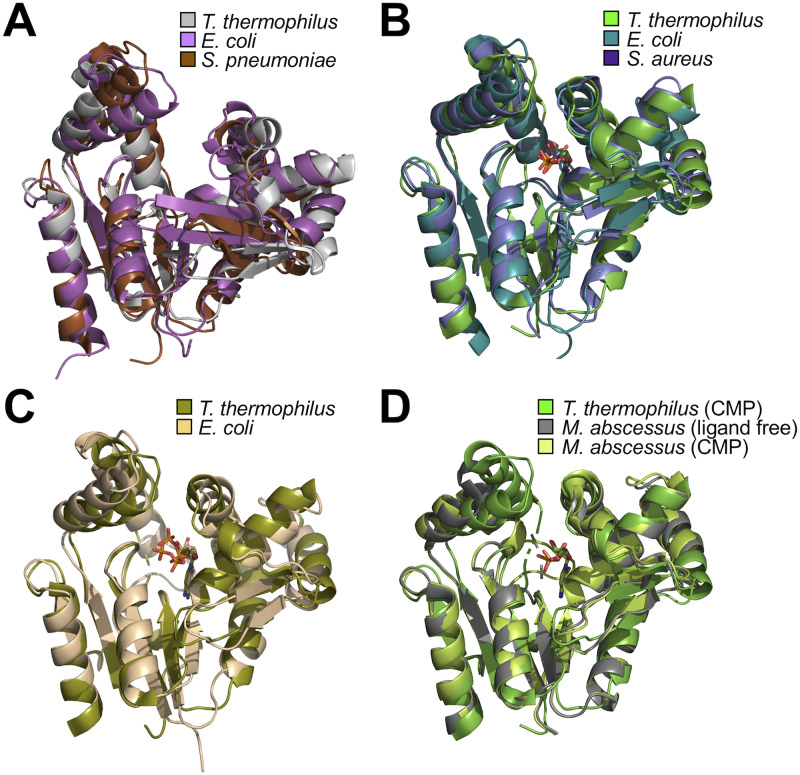
Fig 11. Comparison of CMPK structures.
(A) Ligand-free forms of CMPK from T. thermophilus (gray; PDB code 3W90), E. coli (magenta; 1CKE), and S. pneumoniae (brown; 1Q3T). (B) CMP-bound forms of CMPK from T. thermophilus (green; 3AKE), E. coli (teal; 1KDO), and S. aureus (light purple; 2H92). (C) CDP-bound forms of CMPK from T. thermophilus (olive; 3AKD) and E. coli (light brown; 2CMK). (D) CMP-bound forms of CMPK from T. thermophilus (green; 3AKE) and M. abscessus (light green; 4DIE), and ligand-free form of CMPK from M. abscessus (dark gray; 3R8C). These structures were structurally aligned based on superimposing the backbone structure of their CORE domains. For ttCMPK, the CMP "closed" complex was employed as CMP-bound form.

