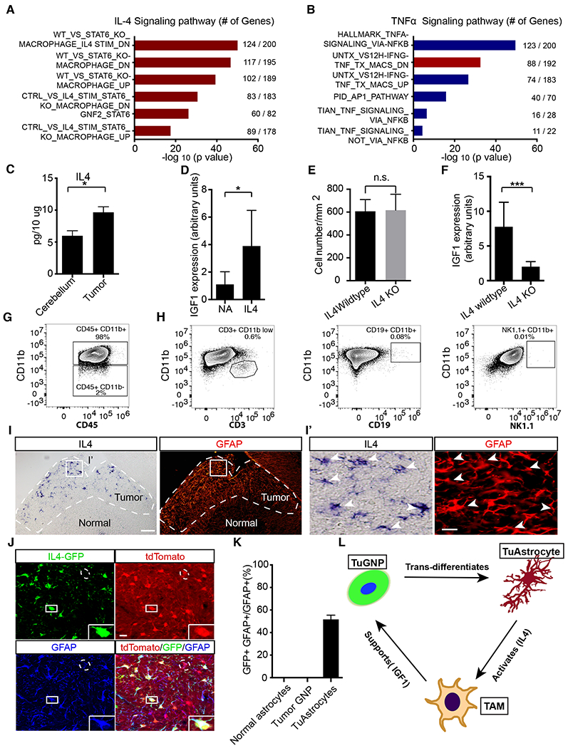
Figure 7. IL4 produced by TuAstrocytes promotes IGF1 expression in TAMs.

A,B, Pathway analysis of differentially expressed genes in TAMs revealed significantly up-regulated signatures associated with IL4 but not TNFa signaling. C, Luminex profiling showed that IL4 level was elevated in the tumor in comparison to normal cerebellum (n=3). D, IL4 stimulation of acutely purified TAMs led to elevated IGF1 level in culture (n=5). E, Quantification of the cell density of TAMs in IL4 wildtype and IL4 KO tumors. F, IL4 knockout led to reduced IGF1 expression level in the tumor mass (n=8 for each genotype). G, Flow cytometry analysis demonstrated that CD11b+ myeloid cells were the dominant population of immune cells in the tumor (n=10). H, Negligible numbers of T, B and NK cells were present (n=4). I-I’, In situ hybridization showed that IL4 was produced by GFAP+ cells in the tumor mass. J, All IL4-GFP+ cells were tdTomato+ and GFAP+, suggesting that IL4 was produced by TuAstrocytes (n=3). K, Quantitative analysis showed that IL4 was expressed by −50% TuAstrocytes but not by normal astrocytes or TuGNPs. L, Working model of an intricately organized TME network in medulloblastoma: a small fraction of tumor GNPs trans-differentiates into TuAstrocytes, which produce IL4 to activate tumor-associated microglia (TAMs), which in turn secrete IGF1 to promote tumor progression.
Scale bar: I =50μm, I′ and J=20μm. Data are Means±SD, Studen t’s t-test; *p<0.05; **p<0.01; ***p<0.001.
