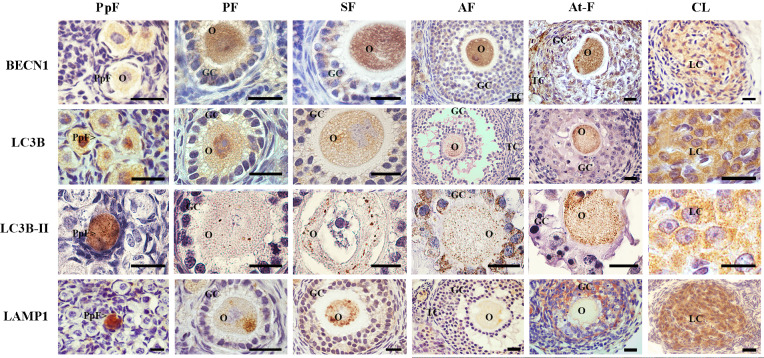
Fig 1. Immunolocalization of autophagy-related proteins in the ovary of L. maximus.
Immunostanning of BECN1, LC3B and LAMP1 in primordial, primary, secondary, antral and atretic follicles and corpora lutea. Note the punctuate pattern of LC3B-II in oocytes of all ovarian structures. PpF: Primordial follicle, PF: primary follicle, SF: secondary follicle, AF: antral follicle, At-F: atretic follicles, CL: Corpora lutea, O: oocyte, GC: granulosa cells, TC: theca cells and LC: luteal cells. Scale bar: 40 μm (short) and 100 μm (large).

