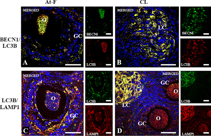
Fig 2. Two-color immunofluorescence of BECN1, LC3B and LAMP1 in atretic follicles and corpus luteum of L. maximus.

Merged immunofluorecense images of BECN1 (cytoplasmic green staining) and LC3B (cytoplasmic red staining) in atretic follicles (A) and corpus luteum (B). Merged immunofluorecense images of LC3B (cytoplasmic green staining) and LAMP1 (cytoplasmic red staining) in atretic follicles (C) and corpus luteum (D). Note the simultaneous expression (citoplasmic yellow staining) of BECN1/LC3B and LC3B/LAMP1 in granulosa cells and oocytes from atretic follicles (A, C) and luteal cells of corpus luteum (B, D). All images show DAPI nuclear staining (blue). O: oocyte, GC: granulosa cells, LC: luteal cells. Scale bar, 60 μm.
