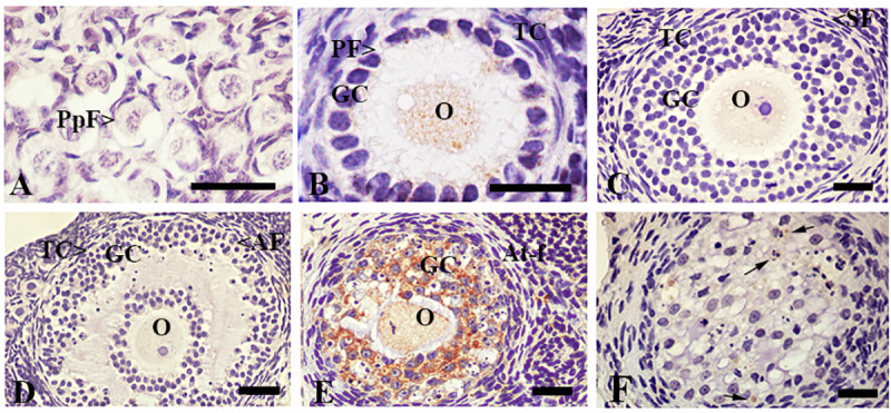
Fig 3. Immunolocalization of ACTIVE CASPASE 3 in the ovary of L. maximus.

Immunostanning of ACTIVE CASPASE 3 in primordial (A), primary (B), secondary (C), antral (D) and atretic follicles (E) and corpora lutea (F). Note that granulosa cells of atretic follicles and luteal cells of corpora lutea showed strong staining for ACTIVE CASPASE 3. PpF: Primordial follicle, PF: primary follicle, SF: secondary follicle, AF: antral follicle, At-F: atretic follicles, CL: Corpora lutea, O: oocyte, GC: granulosa cells, TC: theca cells and LC: luteal cells. Black arrows indicate apoptotic cells. Scale bar: A, B, 40μm; C-F, 100μm.
