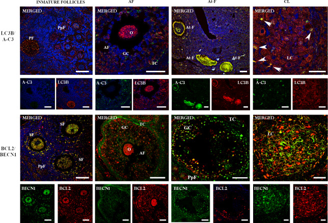
Fig 4. Two-color immunofluorecense of LC3B/ACTIVE CASPASE 3 and BECN1/BCL-2 proteins in the ovary of L. maximus.
Merged immunofluorecense images of LC3B (cytoplasmic red staining) /ACTIVE CASPASE 3 (cytoplasmic and nuclear green staining) and BECN1 (cytoplasmic green staining) / BCL2 (cytoplasmic red staining) in immature, antral and atretic follicles and corpus luteum. Note the simultaneous expression (cytoplasmic yellow staining) of LC3B/ACTIVE CASPASE 3 and BECN1/BCL2 in atretic follicles and immature follicles, respectively. O: oocyte, GC: granulosa cells, TC: theca cells and LC: luteal cells, A-C3: ACTIVE CASPASE 3. White arrows indicate co-localization of A-C3 and LC3B in luteal cells. Scale bar: 40 μm (short) and 60 μm (large).

