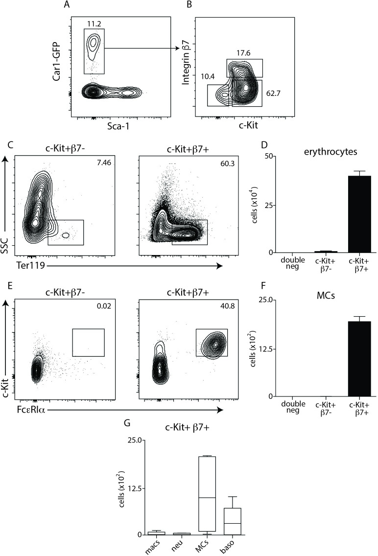
Fig 4. Surface expression of c-Kit and Integrin β7 identify functionally distinct subsets of Car1+ cells.
(A,B), Flow cytometric analysis illustrating expression of c-Kit and integrin β7 on Car1+ cells from the bone marrow. Car1-GFP+ c-Kit+ β7- or Car1-GFP+ c-Kit+ β7+ cells were sort-purified from the bone marrow of mice and seeded into MethoCult and the percentages and total numbers of (C,D) erythrocytes and (E,F) mast cells (MCs) were evaluated by flow cytometric analysis post-culture. (G), The number of macrophages (macs), neutrophils (neu), MCs and basophils (baso) were quantified from Car1-GFP+ c-Kit+ β7+ seeded cultures. (A-G), Representative of at least 3 separate experiments. (G), Illustrates data pooled from 2 separate experiments.

