. 2020 May 3;17(3):237–248. doi: 10.21873/cgp.20184

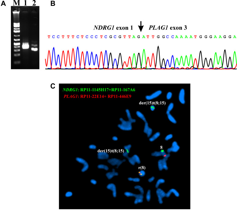
Figure 3. RT-PCR, Sanger sequencing, and FISH analyses of the case 1 of chondroid syringoma. (A) Gel electrophoresis showing the amplified NDRG1-PLAG1 cDNA fragments using the primer combinations NDRG1-1F1/PLAG1-498R1 (lane 1) and NDRG1-23F1/PLAG1-458R1 (lane 2). M, GeneRuler 1 Kb Plus DNA ladder (ThermoFisher Scientific). (B) Partial sequence chromatograms of the cDNA amplified fragment showing the junction position of exon 1 of NDRG1 with exon 3 of PLAG1. (C) FISH analysis on metaphase spreads with PLAG1 probe (red signal) and NDRG1 probe (green signal) showing that the NDRG1-PLAG1 fusion gene was on the ring chromosome 8 (yellow signal). One copy of PLAG1 (red signal) is on chromosome 8. NDRG1 probe (green signal) hybridized also to chromosome 8 and the two der(15)t(8;15) chromosomes.

Figure 3