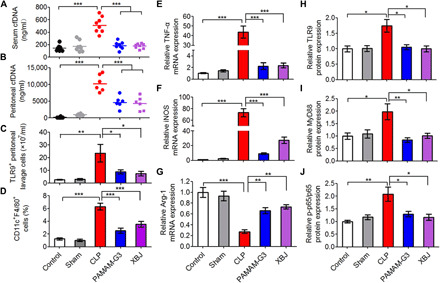
Fig. 2. PAMAM-G3 reverses M1 polarization of peritoneal macrophages through the TLR9-MyD88–NF-κB signaling pathway during severe sepsis.

High-grade CLP was performed on BALB/c mice, followed by intraperitoneal injection of PAMAM-G3 or XBJ (20 mg/kg) 12 hours before and 1 and 12 hours after surgery. (A) Serum and (B) peritoneal cfDNA levels were analyzed after 24 hours after CLP. (C) The number of TLR9+ cells and (D) the percentage of M1-polarized macrophages (CD11c+F4/80+) were assessed in PLF by flow cytometry 8 hours after CLP. Differences were assessed via one-way ANOVA with Tukey’s multiple comparison tests (n = 6 to 8 mice per group; *P < 0.05, **P < 0.01, and ***P < 0.001). The data are expressed as the means ± SEM. (E to J) Peritoneal macrophages were collected 8 hours after CLP, and mRNA was extracted, converted to complementary DNA, and analyzed via real-time polymerase chain reaction (PCR) for (E) TNF-α, (F) iNOS, and (G) Arg-1 gene expression. The data are expressed as fold change relative to the saline-treated normal group and normalized to glyceraldehyde-3-phosphate dehydrogenase (GAPDH) gene expression. In parallel, macrophages were lysed in radioimmunoprecipitation assay (RIPA) buffer before analysis of (H) TLR9, (I) MyD88, and (J) p-p65 protein expression via Western blotting. The data are expressed as fold change relative to the control group and normalized to GAPDH or p65 protein expression. Differences were assessed via one-way ANOVA with Tukey’s multiple comparison tests (n = 5 mice per group; *P < 0.05, **P < 0.01, and ***P < 0.001). The data are expressed as the means ± SEM (n = 3 independent experiments in triplicate).
