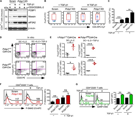
Fig. 5. Increased frequency of FoxP3+ Tregs and enhanced TGF-β signaling in T cells expressing lower PCBP1.

Splenic CD4+CD25− T cells were transduced with scrambled (Scram) or lentiviral shRNA targeting Pcbp1 for 48 hours and cultured in the absence or presence of exogenous TGF-β under iTreg conditions. (A) Immunoblotting of PCBP1 and targets moesin and eIF5A2. β-Actin is shown as loading control. (B and C) FACS analysis (B) and quantification (C) of FoxP3+ T cells 5 days after lentiviral transduction. (D and E) Flow cytometry analyzing CD25-expressing cells among FoxP3+ and FoxP3− T cells (D) and quantification (E) after in vitro culture of splenic CD4+CD25− T cells from Pcbp1f/f and Pcbp1f/fCd4-Cre littermates with allo-DCs in the presence of IL-2 and with or without exogenous TGF-β1 (5 ng/ml). n = 3. (F) Flow cytometry of phospho-Smad2/3 and quantification. (G) Analysis of GARP expression and quantification in scrambled versus Pcbp1 KD T cells. (C and E to G) Error bars represent means ± SD; *P < 0.05, **P < 0.01, and ***P < 0.001 (Student’s t test).
