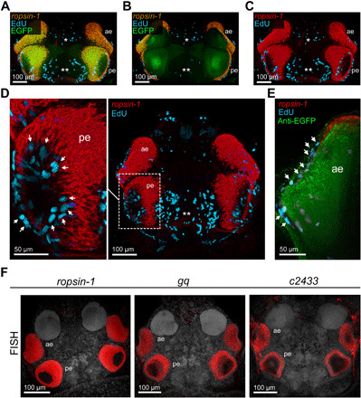
Fig. 6. Retinal growth patterns and molecular signatures of annelid eye photoreceptors.

(A to C) Codetection of incorporated EdU (magenta), riboprobes against ropsin-1 (overlay with EGFP is yellow, and pure signal is red), and EGFP (green) in premature pMos{rops::egfp}vbci2 bristle worms. Single and double asterisks indicate regions of cell proliferation in the anterior and posterior ganglionic region, respectively. (D and E) Similar codetection of EdU (magenta), ropsin-1 (red), and EGFP epitopes (green), including close-ups of the posterior (D, left) and anterior (E) eye region. Arrows point to the proliferative cells in either eye. (F) Fluorescent detection of riboprobes against ropsin-1 (left), gq (middle), and tmdc/c2433 (right) in comparative RNA whole-mount hybridizations, revealing expression of all three genes in eye photoreceptors of the worm head. All dorsal views, anterior to the top. ae, anterior eye; pe, posterior eye.
