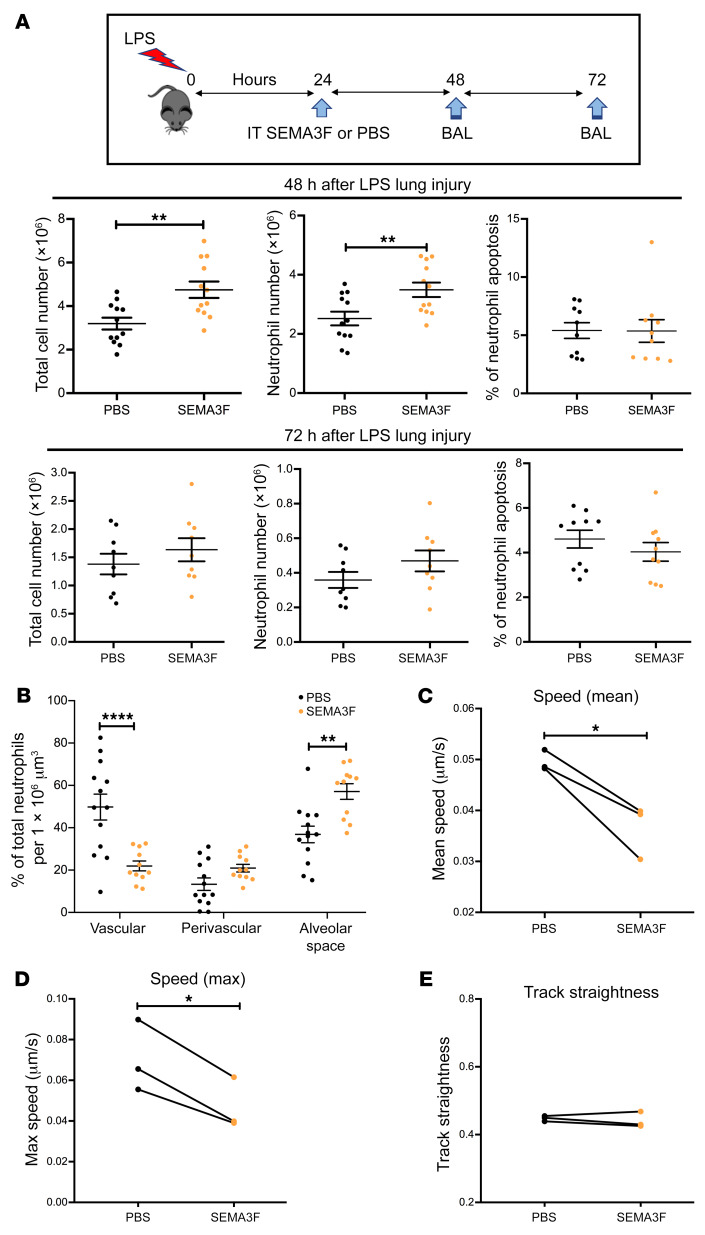
Figure 6. Exogenous SEMA3F retains recruited neutrophils at the injury site in a murine model of acute lung injury.
(A and B) Intratracheal (IT) recombinant SEMA3F (1 μM) was administered to C57BL/6 mice 24 hours after nebulized LPS challenge or PBS. Mice were then sacrificed at 48 and 72 hours and BAL was performed with differential apoptosis cell/neutrophil counts (A), or lungs were retained for fixed lung slice imaging (B). Lungs harvested for lung imaging were instilled with agarose gel, and fixed and stained with the endothelial marker CD31 (green) and the neutrophil marker S100A9 (red). Lungs were imaged by confocal microscopy (Zeiss LSM 880 with Airyscan) with 3D reconstruction and neutrophil position relative to the blood vessels was assigned using Imaris software version 9.1, with at least 80 neutrophils quantified per mouse. Data are mean ± SEM with individual data points from 4 independent experiments (n = 12). (C–E) Naive Catchup (IVM-RED;Lifeact-GFP) mice were sacrificed and lungs were instilled with agarose gel, precision sliced, and imaged by confocal microscopy for 90 minutes with addition of SEMA3F or PBS vehicle control at 30 minutes. Following treatment, neutrophil mean speed (C), maximum speed (D), and track straightness (directionality) (E) were measured and analyzed for 60 minutes using Imaris software version 9.1. Data are from 3 independent experiments (n = 3). Statistical analysis was by 2-way ANOVA and Sidak’s post hoc test (A and B) or paired t test (C–E). *P < 0.05; **P < 0.01; ****P < 0.0001.

