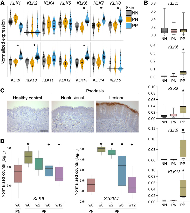
Figure 1. KLK6 expression is significantly elevated in psoriatic lesions and parallels disease activity.
(A) Detection of KLK transcripts by RNA-seq in healthy control skin (NN, gray; N = 90), psoriatic patient nonlesional skin (PN, yellow; N = 26), and psoriatic patient lesional skin (PP, blue; N = 99). P values were computed using Wilcoxon’s rank-sum test; false discovery rate (FDR) was used to control the multiple testing. *P < 0.05 in PP vs. PN. (B) Relative expression of select KLK transcripts by qRT-PCR in a unique cohort of healthy controls and psoriatic patients (N ≥ 6 for all). Mean is indicated by the horizontal line. Box, 25th–75th percentile. Whiskers, minimum and maximum. *P < 0.005 in PP vs. both NN and PN by ordinary 1-way ANOVA with post hoc Tukey’s multiple-comparisons test. (C) Immunostaining of KLK6 in human skin. Results are representative of 4 biological replicates. Scale bar: 100 μm. (D) Detection of KLK6 and S100A7 transcripts by RNA-seq in PN and PP at the indicated weeks’ duration of etanercept therapy in responsive patients (N = 14). Mean is indicated by the horizontal line. Box, 25th–75th percentile. For definition of whiskers and additional details, see Supplemental Methods. *P < 0.05 vs. PN at w0; +P < 0.05 vs. PP at w0 by negative binomial test.

