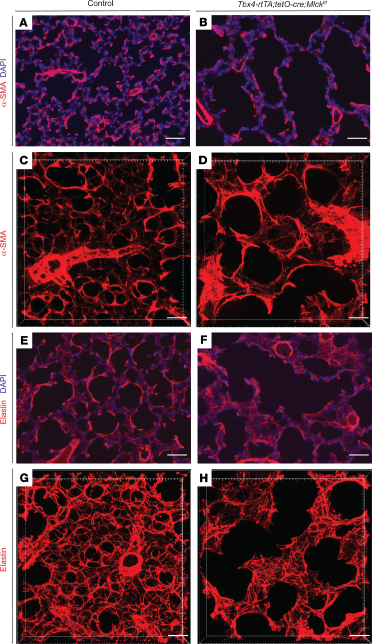
Figure 3. Inactivation of Mlck leads to abnormal myofibroblast and elastin patterns.
(A and B) Representative immunofluorescence staining for α-SMA (red) in sections of the alveolar region showing localization of myofibroblasts on P8. (C and D) Reconstructed 70-μm z-stacks of immunofluorescence staining for α-SMA (red) in the alveolar region of lungs on P8. α-SMA staining in the Mlck-mutant lung was less tightly organized compared with control lung. (E and F) Representative immunofluorescence staining for elastin (red) in sections of the alveolar region on P8. (G and H) Reconstructed 70-μm z-stacks of immunofluorescence staining for elastin (red) in the alveolar region of lungs on P8. Elastin staining in the Mlck-mutant lung was disorganized compared with control lung. Scale bars: 50 μm.

