
Figure 4.
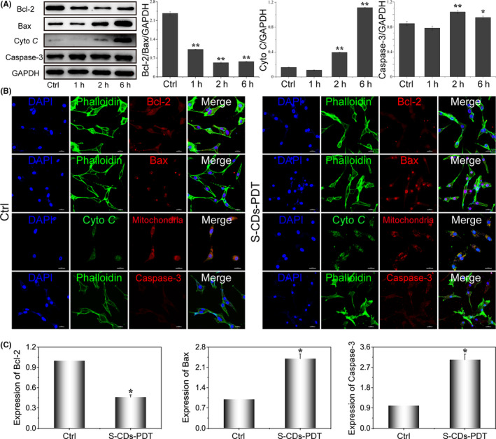
Investigations on the expression of proteins and genes related to cell apoptosis. A, Western blot images and quantitative analysis of apoptosis proteins after PDT mediated by S‐CDs, samples were probed with anti‐GAPDH, anti‐Bcl‐2, anti‐Bax, anti‐Cyto C and anti‐caspase3. GAPDH was used as an internal control. Data are presented as mean ± SD (n = 3). Statistical analysis: *P < .05. **P < .01. B, The immunofluorescence images showing the expression of the essential proteins closely related to apoptosis (Bcl‐2, Bax, Cyto C and caspase3) after S‐CDs mediated PDT. Scale bar: 20 μm. C, Quantitative real‐time PCR analysis of the expression of cell apoptosis‐related genes in U87‐MG after PDT. Data are presented as mean ± SD (n = 3). Statistical analysis: *P < .05. **P < .01