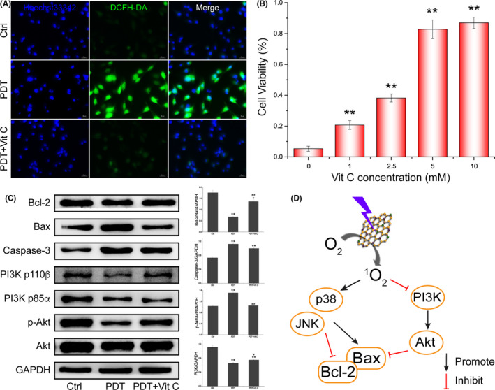
Figure 5.

Detection of ROS and pro‐apoptotic factors in U87‐MG. A, Fluorescent images of ROS generated during S‐CDs‐mediated PDT with a ROS probe, DCFH‐DA. B, Cell viability of U87‐MG after S‐CDs mediated PDT with or without the presence of Vitamin C. C, Western blot images and quantitative analysis of the change of apoptosis‐related proteins and signalling pathway. GAPDH was used as an internal control. Data are presented as mean ± SD (n = 3). Statistical analysis: *P < .05. **P < .01. D, Scheme of signalling pathway changes after S‐CDs mediated PDT
