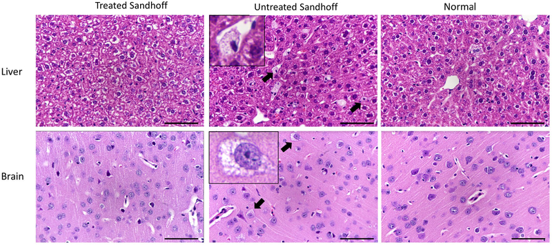
Fig. 5. Histological analysis showed that cellular vacuolation was reduced in the brain and liver of treated Sandhoff mice.
The brain (upper panel) and liver (lower panel) were processed for H&E staining. Treated Sandhoff mice, untreated Sandhoff and normal mice are shown in the left, middle and right columns, respectively. Kupffer cell vacuolation (small, well defined, vesicles with clear to pale-eosinophilic content) in the liver of untreated Sandhoff mice was reduced in treated Sandhoff mice. In the cerebellum, pons, thalamus, hypothalamus and brain cortex of untreated Sandhoff mice, there was neuronal vacuolation, which was significantly reduced in 1 of 3 treated Sandhoff and was not observed in normal mice. Objective x40.

