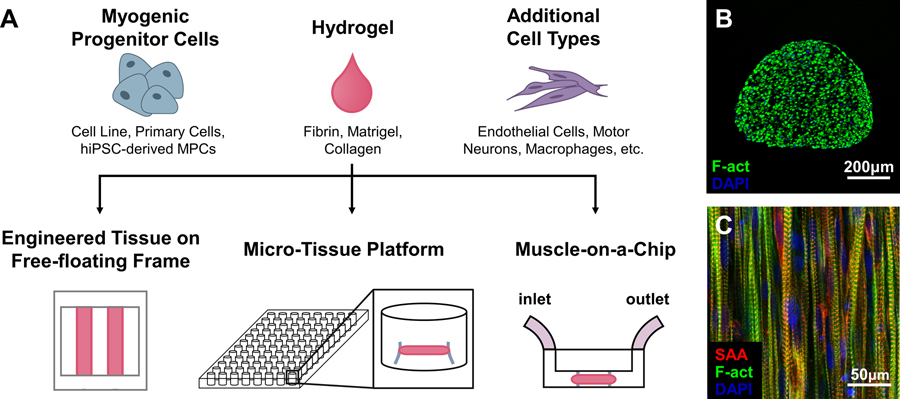
Figure 1.
In vitro tissue-engineered models of human skeletal muscle. (a) Components and platforms for engineering human skeletal muscle tissues for drug testing. Free-floating, dynamic culture improves muscle maturation and enables measurement of force–length relationships; micro-tissue platforms allow for high-throughput drug screening in individual muscle tissues; muscle-on-a-chip platforms have microfluidic feeds for interfacing with other organ-on-a-chip systems. MPC, myogenic progenitor cell. (b-c) Transverse (b) and longitudinal (c) sections of cylindrically shaped engineered human skeletal muscles showing viable myotubes throughout the entirety of the tissue; myotubes are aligned and cross-striated as characteristic of native skeletal muscle. SAA, sarcomeric-actinin; F-actin, filamentous actin; DAPI, nuclei. Tissues in b and c are cultured within free-floating frames on a rocker.

