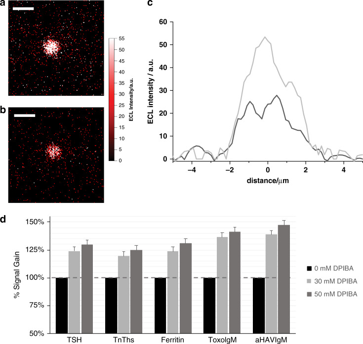
Fig. 4. Bead-based assay and commercial immunoassay using N-dipropyl isobutyl amine (DPIBA).
Electrochemiluminescence (ECL) imaging of a 2.8-μm single bead was obtained by applying a constant potential of 1.4 V (vs. Ag/AgCl) for 4 s in 180 mM tri-n-propylamine (TPrA) and 0.2 M phosphate buffer (PB) with a 50 mM DPIBA and b without DPIBA. Integration time, 4 s; magnification, ×100; Scale bar, 5 μm. c Comparison of the bead profile lines (black, without DPIBA; and gray, with DPIBA). d ECL signal gain observed in the presence of 30 and 50 mM DPIBA in 180 mM TPrA, 0.2 M PB, and 0.1% polidocanol compared with a reference buffer without DPIBA, as measured on a Roche Diagnostics Cobas e 801 immunoassay analyzer using the Elecsys® assays thyroid stimulating hormone (TSH), cardiac troponin T (TnT hs), Ferritin, Toxoplasma gondii IgM (Toxo IgM), and hepatitis A IgM (A‐HAV IgM) and a biomarker containing calibrator as sample. Error bar shows the standard deviation (n = 10).

